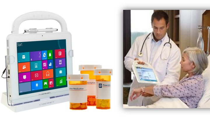
Clinicians are burning out. They are feeling overwhelmed, as they are tasked with more work while at the same time expected to see more patients every day. In a recent survey by the AMA, 54 percent of urologists felt burned out, followed by specialists in neurology (50 percent); nephrology (49 percent); endocrinology, family medicine, and radiology (all at 46 percent). The COVID-19 pandemic worsened things, leaving healthcare groups and hospitals short-staffed to deal with the drastic increase of patients.
Technological aids like electronic medical records (EMR) and medical transcription were supposed to make things easier for clinicians. Unfortunately, they have proven to be a mixed medical bag. This is especially true with EMR, though it’s improving. So it’s understandable why many clinicians view renewed interest in computer aided diagnosis (CAD) with some misgivings. New advances from artificial intelligence and machine learning (especially deep learning) in CAD should hopefully lessen such doubts.
CAD: Potential that Fell Flat
Article Guide
CAD has been around since the mid-1970s. Radiologists were particularly keen about the technology. Computer aided detection (CADe) systems are used to find masses in medical images like a breast or lung for display on a medical monitor. Computer aided diagnosis (CADx) was to help clinicians determine if those displayed masses were benign or malignant. Radiologists hoped both versions would allow them to manage the ever increasing numbers of medical images created by scanning devices quickly and accurately.
Unfortunately, reviews of clinical use of these early systems showed little reduction in radiologists’ workloads. In fact, it did the opposite, increasing them up to 19 percent as the clinicians were forced to dismiss many CAD-positive tags as false while they discovered unflagged or undetected new masses. In 2018, Medicare stopped reimbursements for CAD.
A Case for Computer Aided Diagnosis
In the US, CADe is the only version officially approved by the FDA. That has not stopped development of CADx, though. The benefits of a CAD version that can assist in interpreting an image is too great.
This is especially true in cancer detection. According to the CDC, death by cancer rates in the US have been dropping since 1999. That year, death by cancer was approximately 200 per 100,000 people. That figure dropped to 144 per 100,000 in 2020.
This drop in cancer rates has come at a cost. Many are discovered through the use of screening tests like MRI, CT, X-ray, and ultrasound. Radiologists typically review and interpret the images generated through those tests. Medical equipment dramatically increased the number of images per screening. Some radiologists may have to review 20 to 100 scans with each one containing thousands of images. This is on top of their other duties of seeing patients, performing the screening tests, writing / dictating / editing comprehensive reports of their findings, and discussing said findings with patients and other clinicians. It is no wonder nearly 50 percent of radiologists are experiencing burnout. A CADx program that can interpret medical images with the reliability of an experienced radiologist would greatly speed the process while reducing the workload.
AI and Deep Learning in Computer Aided Diagnosis
Early CAD systems scanned and interpreted medical images using programs written for a specific purpose. A new program had to be written each time clinicians wanted the CAD system to do something different. This limited the system’s abilities especially in diagnosis.
Artificial Intelligence
Previous and current CAD systems like those used for mammograms can only display and examine one image at a time. Using advances in artificial intelligence (AI), future CAD systems can assemble a composite from a variety of images. Depending on the scanning system, they can be from different angles and even taken from previous scans. Even more, scans can be pulled from a variety of sources. These not only include typical scanning devices like MRI, ultrasound, and digital breast tomosynthesis, but even pulled from the EMR stored in a medical tablet. The result is a far more complete composite image with fewer false positives and missed masses.
Deep learning
A subset of machine learning (ML), deep learning (DL) are algorithms that simulate how human beings learn something. CADx systems use it to classify the huge amounts of data they receive from medical scanning devices. Results are already coming in and looking promising. There are CAD systems doing retinal assessment and skin lesion analysis that match human levels in accuracy. Another such system has shown similar accuracy in the detection and determination of prostate cancer from MRI scans. Finally, CAD using DL have both found and determined cancers from CT scans with accuracy equal to thoracic radiologists. Future radiologists will have greater confidence in such CADe and CADx systems and allow them to provide greater patient care.
Closing Thoughts
Computer aided detection and diagnosis (CAD) showed great promise in assisting clinicians in their work, especially in detecting cancer. The system stalled due to limitations in the technology at the time. New CAD systems, using advances in AI and DL, look to finally deliver on that promise as today’s clinicians struggle with burnout. Contact an expert at Cybernet if you’re interested in learning more about all versions of CAD and how they can apply to your medical group.
Join the conversation and connect with us on this and other relevant topics – Follow us Facebook, Twitter, and Linkedin.
4 Ways That AI will Affect Medical Computer Systems
August 28, 2018
The term “artificial intelligence” conjures images straight out of science fiction blockbusters: super-smart machines controlling all aspects of life, and often running wild to destroy their human creators. In reality,…
0 Comments6 Minutes
Prevent Physician Burnout with Health IT That Lifts The Burden
April 5, 2017
EHR can help providers. A lot has been said about how exactly EHR can help everyone in the health care. However, when providers implement EHR the physician productivity and patient satisfaction suddenly drop. The factor…
0 Comments11 Minutes
Implementing Efficient Medication Dispensing Systems through the Use of Medical Tablets and Mobile Solutions
April 6, 2015
Many hospitals and healthcare facilities have increasingly adopted a variety of mobile solutions to address a number of task-related issues. The influx of technological tools like all- in-one computers and medical…
0 Comments4 Minutes
You Can't
Learn from a Pop-up
But we can deliver knowledge to your inbox!
We dive deep in the industry looking for new trends, technology, news, and updates. We're happy to share them with you.
Knowledge, News, and Industry Updates Right in Your Inbox


