Though the concept of digital pathology has been around almost 60 years, it’s only recently that we’ve developed the networks, image resolution, data storage, sharing techniques, and machine learning to truly transform the practice into one of the most important branches of healthcare technology.
But what is digital pathology, and how can cutting-edge data speeds, 4k medical monitors, and brand new digital scanners revolutionize both patient outcomes and clinician workflows?
What is Digital Pathology?
Article Guide
Traditional pathology is the practice of studying body tissue slides and samples, gained from a microscope, for healthcare diagnosis. Of course, that’s not the only definition of the word (which can include the behavior of medical or even social or mental dysfunction), but it’s spot-on for medical purposes.
So, naturally, digital pathology is an extension of the traditional practice, except with more cloud storage. The idea of digital pathology is to scan/import traditional slides of biological tissue into a computer and then to safely and securely share it over the network with other medical professionals.
Digital pathology, to put it succinctly, is an image-based environment for getting, receiving, organizing, and analyzing digital images of glass slides. And because it’s image-based, the whole process needs two important pillars to function: high-definition image scanners and ultra high-resolution medical grade monitors.
Let’s dig into the tech required to pull off digital pathology, both from an input and an output standpoint.
The Technology Required
Digital pathology set-ups aren’t exactly cheap, but they are becoming more commonplace, and more compact. The cornerstone of digital pathology is the digital scanner, a high-end, specialized device that is essentially an upscaled and far more complex version of a scanner for uploading normal pictures in any office or home environment.
However, instead of simply recording and digitizing a flat 2-dimensional image, these pathology scanners take microscopic slides of tissue samples and map their geography down to the cellular level. Obviously, these scanners must be extremely granular: a patient’s life could depend on even the most minute detail being missed.
There are specialized scanners on the market (for only blood samples, or only skin samples), but the FDA has actually recently approved a whole-slide imaging system made by Philips that seems to function with any biopsied tissue. It’s even a compact, all-in-one solution that could be perfect for even small labs.
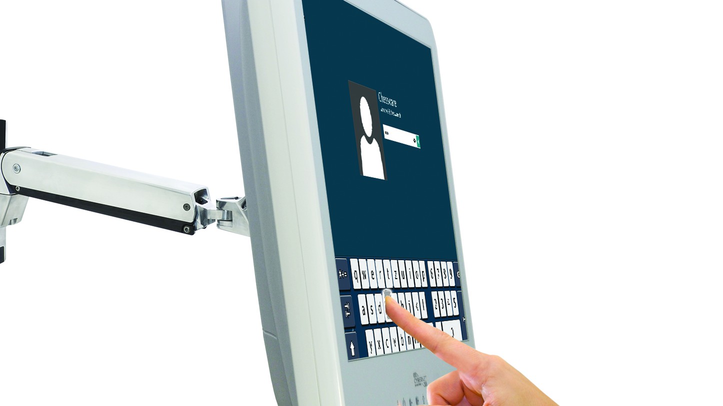
On the end of the equation are high-definition medical monitors for the pathologists viewing the digitized slides. Any specialists called in to analyze the tissue for potentially dangerous biomarkers needs to be confident they won’t miss even a pixel of the high-resolution image being sent to them. Standard monitors in many hospitals are usually chosen for their affordability in large numbers, which works just fine for spreadsheets and internet browsing.
However, a pathologist’s monitor (or any doctor viewing the digital slides) must be a cut above. The days of “1080p” as top-of-the-line are behind us: a monitor for pathology use needs to be 4k, with somewhere in the neighborhood of 8 megapixels. If you’re not familiar with the terms, “4k” refers to a horizontal viewing resolution of 4,000 pixels on the monitor. And 8 megapixels means the monitor can display 8 million pixels. This depth of definition and extreme clarity is absolutely vital for reviewing digital biopsies and making accurate diagnoses.
Where a specialized medical-grade monitor shines is not only it’s high resolution, but in its basic design. An antimicrobial* properties protect the computer casing from deterioration and degradation. Most medical monitors are built from the ground up with durability and a long lifespan in mind: few computers need to run with 24/7 certainty like the computers in a hospital or doctor’s office.
What are the Benefits of Digital Pathology?
Digital pathology can be expensive, not only in the cost of the scanners but in the cost of buying and running enough server space for the ultra-high definition slide images that can take up a lot of hard drive space. And, of course, the infrastructure cost of faster digital download/upload speeds and the hardware to serve those images securely to pathologists across the world.
However, the benefits can’t be underestimated. Machine learning and AI software is improving daily, and can be used for image and pattern recognition to catch problems and help create diagnoses that might have otherwise escaped even the most diligent and experienced pathologist. For instance, the FlexMIm project can analyze 3.5 million pixels per quad-core processor per minute. That’s a rate even the five fastest pathologists working together couldn’t touch.
This pattern recognition can even be compared, by the program or the pathologist, with historical data and images captured from all around the world. This creates a massive knowledge base that only expands with more users, and subsequently becomes even more accurate with each pathologist who uses it.
What Does Digital Pathology Do for Patients?
Perhaps the most important element of digital pathology, at least to the patients, is the increased speed of diagnosis.
Pathologists often seek a second opinion, to run their results by a colleague to ensure accuracy. In the past, this would involve sending physical microscope slides across country (or even internationally!) depending on the location of the original pathologist and the colleague they’re seeking out. While this is going on, the patient is waiting on tenterhooks to learn if their tumor is really benign, or if they have a serious blood disease, cancer, or a million other possible real or imagined maladies. This can take days or weeks while the samples travel, and while the pathologist double checks the work and sends it all back.
With digital pathology, a patient doesn’t have to agonize for days about their potential condition. Instead, the original pathologist can send the highly-detailed digital slides to other pathologists or specialists in the relevant field over the internet. Not only does this increase the speed of diagnosis and lower wait times, but it also means that geography is no longer a limiting factor.
If the expert is on the other side of the globe or down the street it makes no difference, which greatly expands the pool of potential experts for any given patient and vastly increases their odds of a more accurate diagnosis and a better outcome in the long run.
The Right Tools for the Job
There’s a saying in computer programming that works really well for digital pathology: “garbage in, garbage out.” A pathologist reviewing a digital biopsy either right on site or remotely can only be as accurate as the original scan and the monitor they’re viewing it on.
It may cost a little extra to get a high-quality pathology scanner and a UHD medical monitor, but digital pathology is practically impossible (or at least inaccurate) without both.
To learn more about the medical monitors and equipment necessary to make the most of a digital pathology image, contact Cybernet today.
Medical-Grade vs Regular Monitors: Differences Explained
August 7, 2018
Computer monitors have become an integral part of our lives, not only at home and work, but in every facet of society today. You see them at the grocery store checkout line when the clerks scan your food, at the gas…
0 Comments7 Minutes
How Medical Tablets Barcode Scanners Are Reducing Errors in Healthcare
December 3, 2015
Technology is dramatically changing health care as we know it. Patient care is improving. The speed in which medical decisions are being made about a patient’s care is quicker than ever. Electronic medical record (EMR)…
0 Comments4 Minutes
You Can't
Learn from a Pop-up
But we can deliver knowledge to your inbox!
We dive deep in the industry looking for new trends, technology, news, and updates. We're happy to share them with you.
Knowledge, News, and Industry Updates Right in Your Inbox


